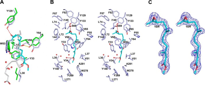
Figure 6.
Ligand binding interactions in mJHBP and salivary D7. A, stick diagrams of conserved residues in the binding pockets of mJHBP (carbon atoms colored light blue) and An. stephensi salivary D7 protein AnStD7L1 (carbon atoms colored green). The JH III ligand in mJHBP is colored cyan with oxygen in red, and the U46619 ligand in AnStD7L1 is colored light gray, with oxygen colored red. The hydrogen bond between Tyr-129 of mJHBP and the JH III epoxide group is shown as a red dashed line, and the hydrogen bond between the equivalent of Tyr 64 in AnStD7L1 and the ω-6 hydroxyl is shown as a yellow dashed line. Numbering of the residues Is for mJHBP. B, stereoview of mJHBP binding pocket with JH III (cyan) bound. Hydrogen bonds are shown as red dashed lines, and ordered water molecules in the binding pocket are shown as red spheres. C, stereoview of simulated annealing omit Fo − Fc electron density (contoured at 3 σ) with the final JH III ligand model inserted. The map was calculated with all JH III ligand coordinates omitted prior to simulated annealing refinement in Phenix.refine (35). The C-10 position having an R configuration is labeled.

