Figure 7.
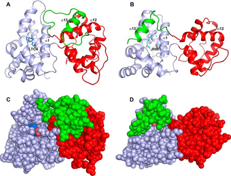
Structural comparison of a salivary D7 protein (AnStD7L1) and mJHBP. A, ribbon diagram of AnStD7L1 with the N-terminal domain colored light blue, the C-terminal domain colored red, and the extreme C terminus including helix α13 colored green. The leukotriene C4 ligand (LTC4) is shown in stick representation with carbon colored blue and oxygen red. B, mJHBP oriented and colored as in panel A with the JH III ligand shown in stick representation with carbon colored cyan and oxygen red. In the case of mJHBP, the C-terminal portion including helix α13 is positioned to cover the entry to the binding pocket. C, space filling representation of panel A to show the open entry to the N-terminal domain binding pocket that accommodates the 20-carbon fatty acid of leukotriene C4. D, the space filling model of mJHBP, oriented as in panel B, shows nearly complete burial of the bound ligand and occlusion of the entry channel.

