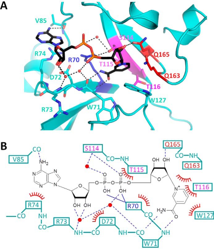
Figure 2.

Interaction between catalytic domain and βNAD+ molecule. A, close-up views of the βNAD+ binding pockets in βNAD+-bound pierisin(1–233)E165Q. The catalytic domain is colored cyan. The (Q/E)XE motif, STS motif, and conserved Arg residue are colored in the same manner as in Fig. 1C. Water molecules are represented as red spheres. Hydrogen bonds are represented by dashed black lines. βNAD+ molecules are depicted as stick models. B, schematic drawing of the interaction between βNAD+ and pierisin(1–233)E165Q. Solid and dashed blue lines represent ionic interactions and hydrogen bonds, respectively. Red arcs with spokes represent van der Waals interactions or π-π interactions.
