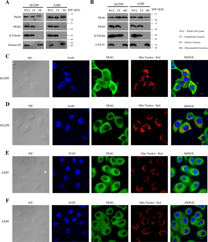
Figure 3.
Subcellular localization of PKM isoforms and their validation. A, immunoblots of PKM1 and PKM2 from the lysate of H1299 (left panel) and A549 (right panel) cells collected by fractionating the cytoplasm and the nucleus (WCL, whole cell lysate; CF, cytoplasmic fraction; and NF, nuclear fraction). PARP and β-tubulin served as loading controls for nucleus, and cytoplasm respectively. B, immunoblots of PKM1 and PKM2 from lysate of H1299 (left panel) and A549 (right panel) cells collected by fractionating the cytoplasm and the mitochondria (WCL, whole cell lysate; CF, cytoplasmic fraction; MF, mitochondrial fraction). COX IV and β-tubulin served as loading controls for mitochondria and cytoplasm, respectively. C–F, confocal microscopy images demonstrating the subcellular localization of PKM1 or PKM2 in H1299 and A549 cells, immunostained with antibodies of PKM2 (green) or PKM1 (green), mitochondria stained with Mito-tracker Red (red) and the nucleus was stained with DAPI (blue). Merged figures are shown with a scale bar of 20 μm.

