Figure 7.
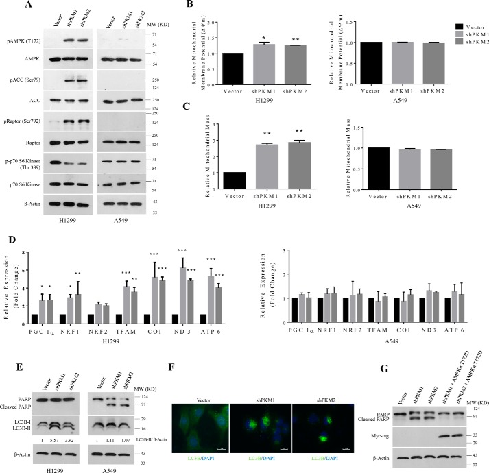
AMPK signaling reprograms energy metabolism pathway to sustain energy homeostasis and to prevent apoptotic cell death. A, immunoblots from the protein lysate of H1299 (left panel) and A549 (right panel) cells stably transduced with lentivirus containing control vector (pLKO.1), shPKM1, or shPKM2, to show AMPK signaling activation. B and C, bar diagram depicts the relative mitochondrial membrane potential (B) and mitochondrial mass (C) in H1299 (left panels) and A549 (right panels) cells stably transduced with control vector (pLKO.1), shPKM1, or shPKM2; with statistical analysis (where n ≥ 3; mean ± S.D.), and the level of significance was tested using unpaired Student's t test. *, p < 0.05; **, p < 0.01. D, quantitative RT-PCR analysis to show the relative expression change of genes involved in the mitochondrial biogenesis (PGC 1α, NRF1, NRF2, and TFAM) and mitochondrial-encoded subunits of electron transport chain complexes (COX 1, ND3, and ATP6) from H1299 (left) and A549 (right) cells for stable PKM1 and PKM2 knockdown. The bars represent the -fold change after normalizing with the control of each group (vector transfected); with statistical analysis (where n ≥ 3; mean ± S.D.) and the level of significance was tested using two-way analysis of variance with Tukey's multiple comparisons test. *, p < 0.05; **, p < 0.01; ***, p < 0.001. E, immunoblots from the protein lysate of H1299 (left panel) and A549 (right panel) cells transduced with lentivirus containing empty vector (pLKO.1), shPKM1, or shPKM2 to measure autophagy and apoptosis using LC3B-II and cleaved PARP as markers. F, confocal microscopy images depicting the autophagic puncta formation in H1299 cells stably transduced with shPKM1 and shPKM2, immunostained with LC3B antibody and secondary anti-Alexa Fluor 488. Nucleus was stained with DAPI (blue). Merged figures are shown with a scale bar of 20 μm. G, immunoblot from the protein lysate of A549 cells, transduced with lentivirus containing empty vector (pLKO.1), shPKM1, shPKM2, and, in addition, ectopic expression of Myc-tagged AMPKα2 constitutively active T172D mutant (AMPKα2T172D-Myc) in cells knocked down for PKM1 and PKM2 probed with Myc-tag and PARP antibodies to validate the expression of AMPKα2T172D-Myc and to measure apoptosis.

