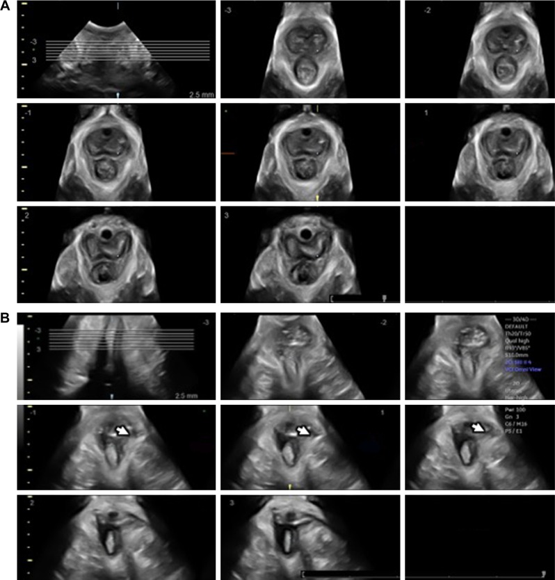
Figure 3.
Tomographic ultrasound imaging of the levator ani muscle obtained by three-dimensional/four-dimensional transperineal ultrasound.
Notes: The transducer is placed in the midsagittal plane. (A) Findings are normal, with no avulsions noted. (B) An avulsion of the muscle (arrow heads) is shown.

