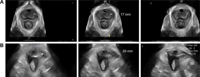
Figure 4.
Measurement of the levator–urethra gap (LUG) on tomographic ultrasound imaging (TUI).
Notes: The images show the three central TUI slices used for determination of the LUG in a normal patient (the upper three images, A) and in a patient with unilateral avulsion (lower three images, B). The patient’s left side is shown on the right in all images. All measurements in (A) are normal and <25 mm. Conversely, there is an obvious left-sided levator avulsion injury visible in (B) with measurements >25 mm. LUG should be measured on both sides for all three central images, but for clarity reasons in the images, only one LUG is measured on one side for each woman.

