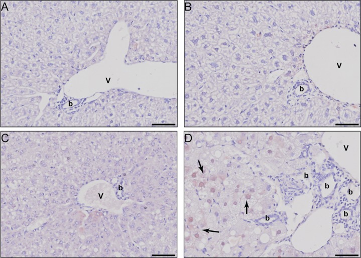
Fig 3. Immunohistochemical evaluation of hepatic AFP.
Representative photomicrographs for AFP stained liver sections of (A) sesame oil vehicle treated females, (B) sesame oil vehicle treated males, (C) 30 μg/kg TCDD treated females, and (D) 30 μg/kg TCDD treated males. Scale bar represents 50 μm. The portal vein is designated by the letter V, bile ducts with the letter b, and AFP positive stained regions by solid black arrows.

