
Figure 3. In vivo.
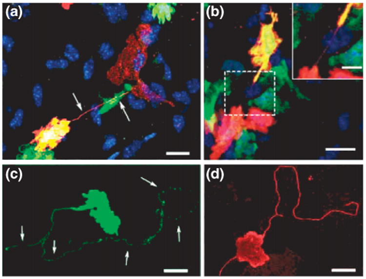
intercellular nanotubes (ICNs) observed between cells in the corneal stroma tissue of mice. (a), Chimeric mouse corneal whole mount reveals a donor-derived (green fluorescent protein (GFP)+ /green) major histocompatibility complex (MHC) class II+ (red) and double positive (yellow) cell connected via a fine ICN (arrows) to a resident MHC class II+ GFP- cell (red only). (b), Two donor-derived MHC class II+ cells expressing varying amounts of GFP joined by a fine, straight ICN. (c) and (d), Long, nonbridging membrane nanotubes on MHC class II+ cells in the naive, (c), and inflamed, (d), mouse corneal stroma. Scale bar, 20 μm; inset scale bar, 10 μm. (Reprinted with permission from Ref 19. Copyright 2008 AAI).