
Figure 6.
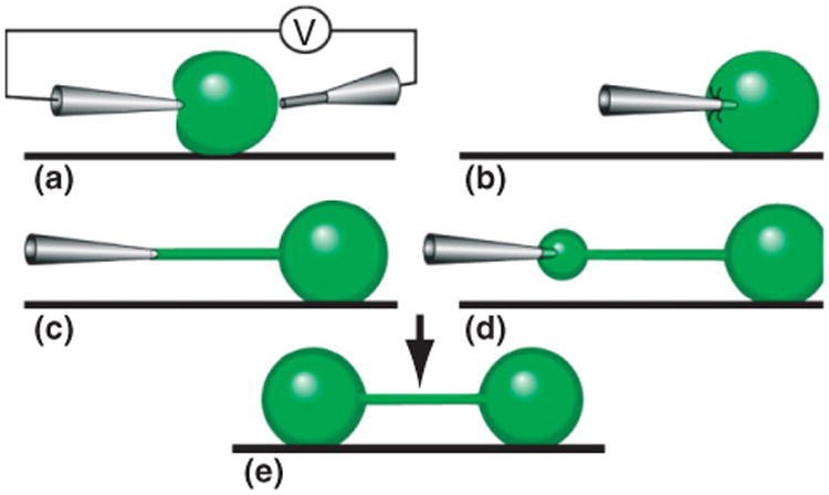
Schematic illustration of the nanotube-vesicle network fabrication principle. (a), By a combination of mechanical deformation and electric pulses across the liposome, a microinjection needle is inserted into a unilamellar liposome connected to a multilamellar protrusion (not shown). (b), After the lipid has adhered to the injection needle, the micropipette is pulled away from the liposome. (c), A lipid nanotube is created between the tip of the micropipette and the liposome. (d), By applying a low pressure in the microinjection needle, ejected liquid expands the nanotube into a liposome at the microinjection tip, transferring additional lipid material from the multilamellar liposome (not shown). (e), After the liposome has reached a desired size, it is allowed to adhere to the surface. Thereafter, the micropipette is removed by applying electric pulses and simultaneously pulling it out of the liposome.