
Figure 8.
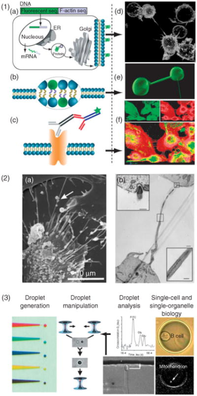
Materials and methods of nanotubular studies. (1) Fluorescent imaging techniques such as confocal imaging provide spatiotemporal information based on fluorescent staining. Labeling of cells is based on, (a), genetically modified cell lines expressing fluorescent proteins, (b), membrane stains, or, (c), antibodies. The panels to the right give specific examples of nanotubes tagged using these methods. (d), B-cells expressing glycosylphosphatidylinositol anchored GFP. (e), nanotube-vesicle network stained with the membrane dye DiO. (f), Primary macrophages stained with phalloidin for F-actin (red) and with monoclonal antibody against α-tubulin (green). (2) Electron microscopy provides structural details on a nanometer resolution scale. (a), Scanning electron microscopy can provide external structural information. Adapted from Veranic et al.51 © 2008, with permission from Elsevier. (b), Transmission electron microscopy illuminates the internal structure, such as membrane junctions. (Adapted with permission from Ref 51. Copyright 2008 Nature Publishing Group). (3) Femtoliter- and picoliter-sized droplets as nanolaboratories for manipulating single cells and subcellular compartments is a potential route to analyze cellular nanotubes. A potential route to analyze intercellular nanotubes could be the use of droplet-nanolaboratories as has been demonstrated for single cells and subcellular compartments. (Reprinted with permission from Ref 81. Copyright 2009 ACS).