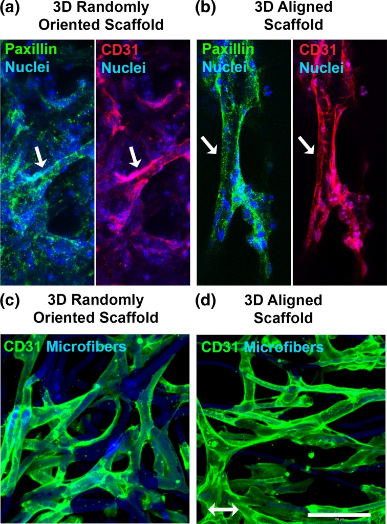
Figure 8.
iPSC-EC morphology and focal adhesion assembly in 3D microfibrous scaffolds. (a–b) Confocal microscopy images depict paxillin expression in iPSC-ECs within 3D randomly oriented (a) or aligned (b) microfibrous scaffolds. CD31 (red), paxillin (green), and nuclei (blue). Arrows point to a representative CD31+ iPSC-EC. (c–d) Confocal microscopy images of iPSC-ECs cultured within randomly oriented or aligned scaffold depict elongated cellular morphology and wrapping of cell bodies around microfibers. The iPSC-ECs were visualized by CD31 (green) expression. The microfibers were visualized by autofluorescence (blue) near the 480 nm emission wavelength. Arrow denotes bulk microfiber alignment direction. Scale bar: 100 µm.

