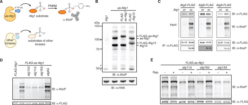
Figure 3. A chemical genetic assay for Atg1 kinase activity.
(A) Schematic of the chemical genetic strategy for monitoring Atg1-dependent thiophosphorylation. Analog-sensitive (as) Atg1 can accept A*TPγS, an N6-substituted ATPγS analog, to thiophosphorylate its substrates while other kinases reject the analog. Following alkylation with para-nitrobenzyl mesylate (PNBM), labeled substrates are immunodetected with anti-thiophosphate ester (thioP) antibody.
(B) Extracts derived from logarithmically growing cells with indicated genotypes were treated as diagrammed in part (A) and analyzed by SDS-PAGE (gel system 1; see Experimental Procedures for details) and immunoblotting (IB) with indicated antibodies. HXK, hexokinase; *, non-specific band.
(C) The indicated extracts were treated as diagrammed in part (A) and subjected to immunoprecipitation (IP) with anti-FLAG agarose resin. Eluates and extract (input) samples were resolved by SDS-PAGE (gel system 2; see Experimental Procedures for details) followed by immunoblotting (IB) with indicated antibodies. wt, wild-type.
(D) Extracts derived from logarithmically growing cells with indicated genotypes were treated as diagrammed in part (A) in triplicate (one of which is shown) and analyzed by SDS-PAGE (gel system 2) and immunoblotting (IB). For quantitation and statistics, see Figure S3F.
(E) Logarithmically growing cells with indicated genotypes were treated with rapamycin (Rap.) or mock treated prior to analysis as in part (D). Dotted line indicates splicing of gel-image data from the same gel. For quantitation and statistics, see Figure S3G.
See also Figure S3.

