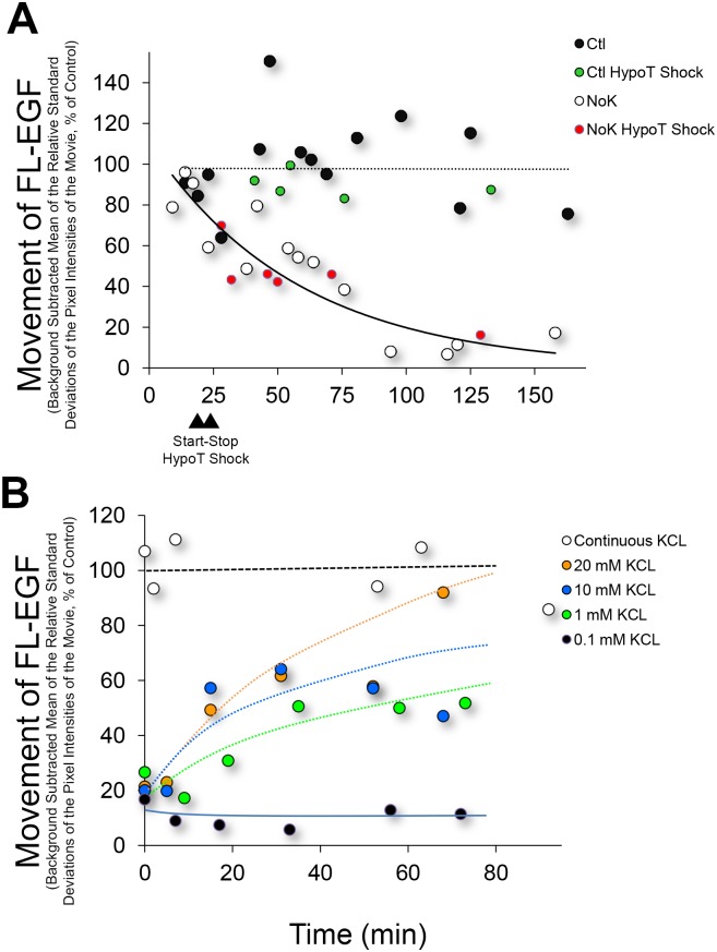
Fig 2. Time course for inhibition of FL-EGF movement and rescue of inhibition of movement by addition of KCl.
(A) Huh-7 cells were exposed to FL-EGF, Hoechst, and washed and incubated in control (Ctl, black circles) or K+ free (NoK, white circles) live medium, and imaged at the times indicated. FL-EGF movement was quantified by measuring the mean of the relative standard deviations of the movie pixel intensities and normalizing to the mean value of the controls. Each circle represents a field of cells containing on average 143 nuclei for these experiments. Movement of FL-EGF containing vesicles is seen to decrease through approximately 90 min, whereupon it plateaus near 10% of control. Exposure of cells to hypotonic shock for 5 min (stop and start times indicated by triangles) did not appear to affect the time required to reduce movement under K+ free medium (red circles) nor affect control movement (green circles) following re-addition of isotonic medium. (B) Cells were exposed to Ctl (white circles) or NoK (black and colored circles) medium for 90 min followed by addition of KCl at the concentration indicated.

