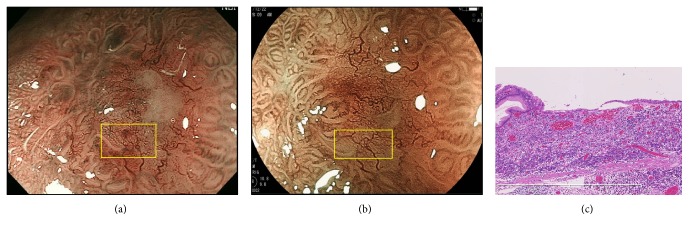
Figure 5.
A signet-ring cell carcinoma in group C. (a) MSP was absent on ME-NBI (yellow box). (b) MSP was also absent on ME-BLI (yellow box). (c) Histological view of the yellow box in Figures 5(a) and 5(b). The glandular architecture was destroyed due to infiltration of signet-ring cell carcinoma in the entire mucosal layer.

