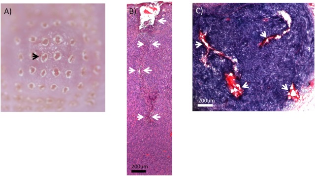
Fig 1. Ablative fractional photothermolysis.
(A) photo of skin surface appearance immediately after ablative fractional photothermolysis (100 mJ pulse energy). Black arrow indicates one of the laser-generated holes formed by tissue vaporization/ablation. There is an absence of graying or blistering immediately after laser exposure, which also suggests an absence of major thermal injury. (B) H&E-stained CT26.CL25 tumor immediately after the ablative fractional photothermolysis (aFP) laser treatment with a pulse energy of 100mJ. White arrows indicate an ablated hole which is characteristic of aFP procedures. The ablated hole appeared to be collapsed and distorted within the tumor tissue. (C) CT26.CL25 tumor immediately after aFP, stained by NBTC staining that shows vital cells as a blue color. White arrows indicate dead cells caused by physical effects of the laser treatment. Most of the tissue within the aFP volumes exhibited a blue staining, indicating an absence of widespread thermal injury or tissue bulk heating.

