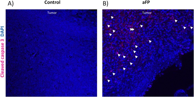
Fig 5. Immunohistochemical staining for apoptotic tumor cells.
(A and B) Immunohistochemical staining for apoptotic cells in the tumor 1 day after aFP in the control group and aFP group respectively. Representative images are shown. Cells stained as red color, which are indicated by white arrow heads are apoptotic cells.

