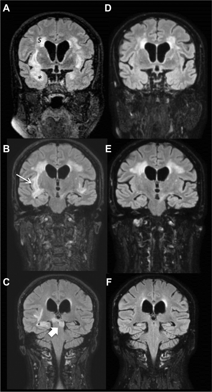
Figure 2.

Brain MRI images before and after B-cell depleting therapy.
Notes: Brain MRI images from February 2016 showed extensive and bilateral focal or confluents subcortical and deep WM lesions, hyperintense in long TR sequences, involving (A) semioval centers (S), temporal lobe (*), (B) external capsule, claustrum and subinsular regions (thin arrow) and (C) midbrain (arrow) without contrast enhancement. Moderate subcortical atrophy with dilation of lateral ventricles. Brain MRI in September 2016, 6 months after RTX treatment, showed reductions in numbers and size of hyperintensity lesions in WM, especially in temporal lobes bilaterally (D and E) and brainstem (F).
Abbreviations: MRI, magnetic resonance imaging; WM, white matter; RTX, rituximab.
