
Figure 3.
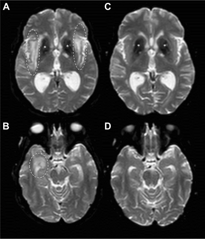
DWI images before and after B-cell depleting therapy.
Notes: Brain MRI DWI sequences from February 2016 (A and B) showed hyperintensity in right temporal lobe (dashed circles), external capsule and claustrum (dashed ovals) with increased apparent diffusion coefficient (not shown) suggesting vasogenic edema. *Basal ganglia calcification. On September 2016, 6 months after RTX treatment, brain MRI DWI sequences (C and D) showed complete disappearance of edema.
Abbreviations: MRI, magnetic resonance imaging; DWI, diffusion-weighted imaging; RTX, rituximab.