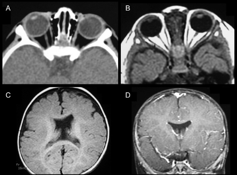
Fig. 1.

Bilateral optic nerve hypoplasia (ONH) with agenesis of septum pellucidum on neuroimaging (Case 1). (A) Computer tomography showed atrophy of the right optic nerve. (B) T1-weighted magnetic resonance imaging (MRI) revealed atrophy of bilateral optic nerves. Absent septum pellucidum was demonstrated on (C) axial view (T1-weighted MRI) and (D) coronal view (T1-weighted contrast MRI).
