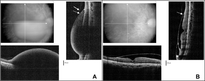
Fig. 2.
Optical coherence tomography (OCT) findings before and after Nd:YAG laser membranotomy in Case 1. (A) The very faint overlying membrane is the posterior hyaloid (solid arrow), and on the retina aspect is the more reflective internal limiting membrane (ILM) (dotted arrow). The infrared image shows the location of the vertical and horizontal spectral domain-OCT (SD-OCT) scan area. (B) One week after Nd:YAG laser membranotomy, the detached ILM (dotted arrow) was present and residual blood was found in the subhyaloidal space (asterisk) in the lower part.

