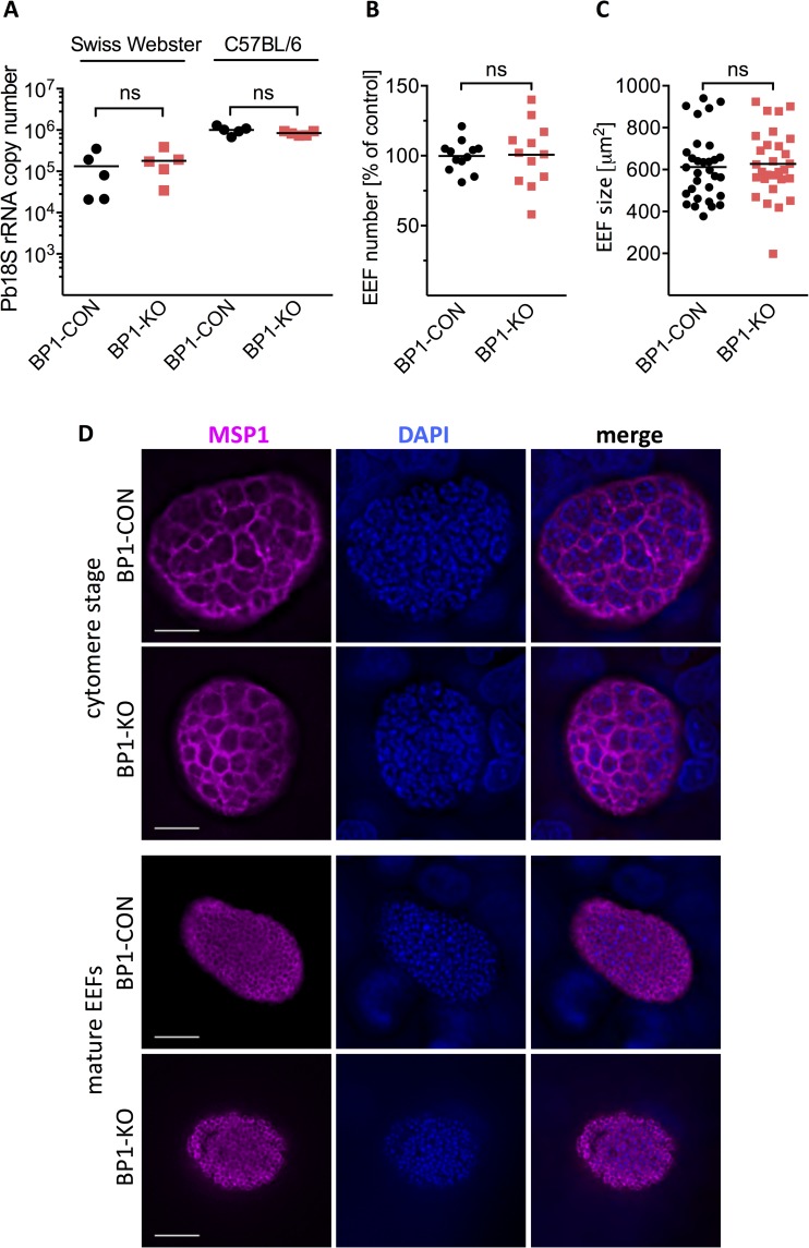
Fig 2. Normal in vivo and in vitro growth of berghepain-1 knockout liver stage parasites.
A. Development of berghepain-1 knockout liver stage parasites in vivo. 10,000 BP1-CON or BP1-KO sporozoites were injected i.v. into Swiss Webster or C57BL/6 mice. RT-qPCR analysis of liver RNA 40 h after infection found no significant reduction of BP1-KO liver stage growth as measured by parasite 18S rRNA copy number. Results from one representative experiment using BP1-CON and BP1-KO clone 1, from a total of three experiments, is shown. B. & C. Development and size of berghepain-1 knockout EEFs in vitro. HepG2 cells were infected with BP1-CON and BP1-KO parasites and at 60 h post infection the number of EEFs was counted (B) and their sizes measured (C). Pooled results of two independent experiments, one using BP1-KO clone 1 and one using BP1-KO clone 2 are shown. D. Immunofluorescene assays of late liver stage berghepain-1 knockout parasites. HepG2 cells infected with either BP1-CON or BP1-KO clone 1 sporozoites were fixed 56 h (cytomere stage) and 72 h (mature EEF) post-infection. EEFs were stained for MSP1 to visualize the parasite membrane (magenta) and DNA was stained with DAPI (blue). Scale bars: 10 μm.

