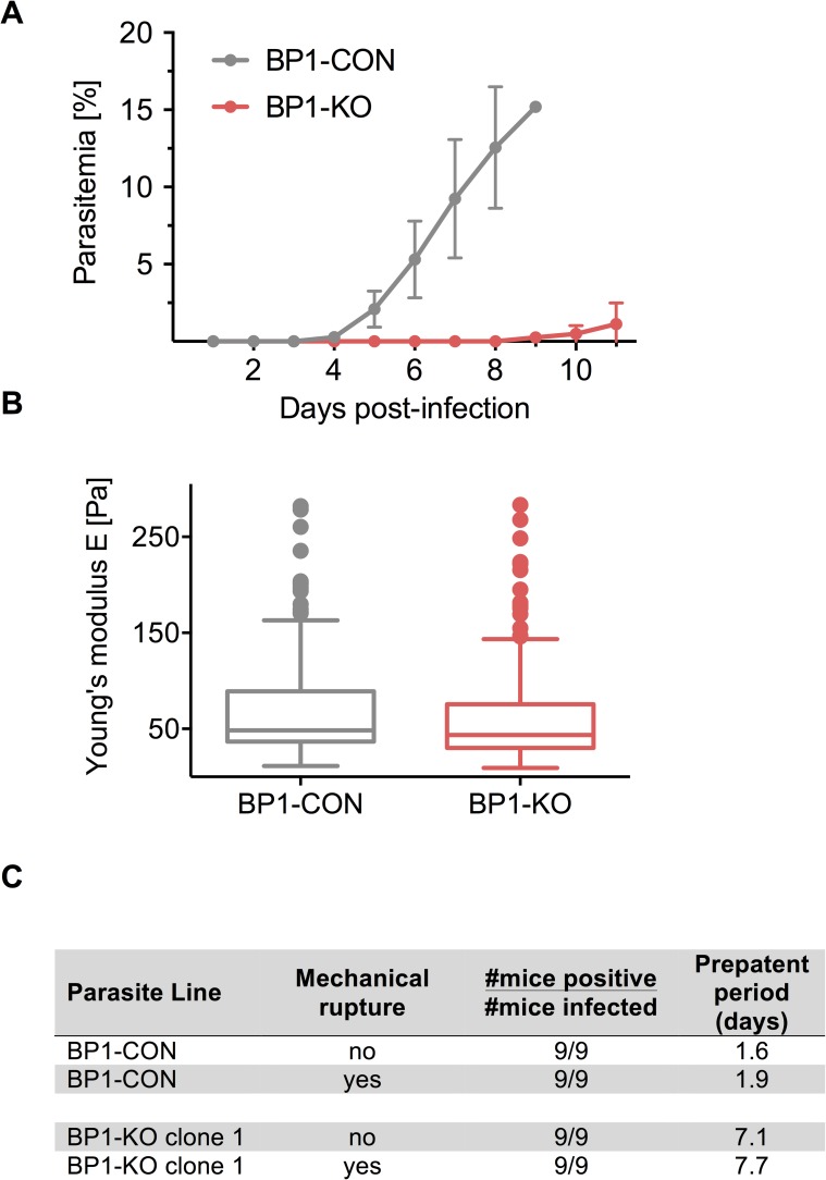
Fig 4. Berghepain-1 knockout merosomes have reduced infectivity.
A. Detection of blood stage parasitemia following injection of merosomes. Five merosomes of BP1-KO or BP1-CON parasites were injected i.v. per mouse and parasitemia was monitored daily for 11 days. Results from two representative experiments are shown and the average daily parasitemia ± SD of 5 mice infected with BP1-CON and 6 mice infected with BP1-KO clone 2 are plotted. B. Atomic force microscopy of the merosome membrane. Merosomes of BP1-CON and BP1-KO parasites were fixed and subject to AFM to measure Young’s modulus (E). Cell elasticity was measured on one point of each cell adhered to a poly-L-lysine-coated glass slide with 5 force-distance curves per cell. Data is displayed in a Tukey box and whisker plot, with a horizontal line representing the median and individually plotted outlier values. Shown are pooled values of seven independent experiments, four performed with BP1-KO clone 1 and three with BP1-KO clone 2, corresponding to approximately 195 cells for each clone. C. Detection of blood stage parasitemia following injection of unruptured or mechanically ruptured merosomes. 5000 ruptured or unruptured merosomes of BP1-KO or BP1-CON parasites were injected i.v. per Swiss Webster mouse. Average prepatent period of BP1-CON and BP1-KO clone 1 from two independent experiments performed are shown.

