Figure 3.
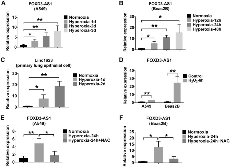
Oxidative stress or hyperoxia induces FOXD3-AS1 expression in lung epithelial cells. A–C) A549, Beas2B, and primary lung epithelial cells were exposed to the hyperoxia for the indicated times. The expressions of FOXD3-AS1 in A549 (A), Beas2B (B), and primary lung epithelial (C) cells were determined using real-time PCR. D) A549 and Beas2B cells were treated with 90 µM H2O2 for 6 h. The expression level of FOXD3-AS1 was measured using real-time PCR. E, F) A549 (E) and Beas2B (F) cells were exposed to normoxia or hyperoxia for 24 h in the presence or absence of 5 mM NAC. Real-time PCR was performed to determine the level of FOXD3-AS1. Data represent 3 independent experiments. *P < 0.05, **P < 0.01.

