Figure 5.
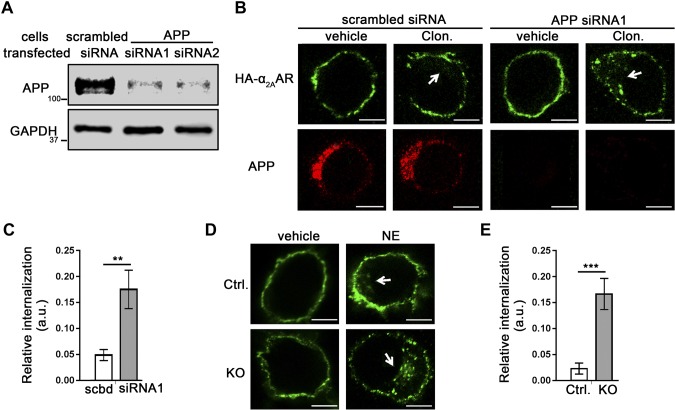
Loss of endogenous APP in N2a cells increases α2AAR internalization. A–C) N2a cells stably expressing HA-α2AAR were transfected with APP siRNAs or a scrambled siRNA. Internalization of surface HA-α2AAR was examined by an Ab-prelabeling method. A) Western blot image showing expression of the knockdown efficiency of scrambled or APP siRNAs. B) Representative images showing internalization of cell surface HA-α2AAR after 1 µM clonidine treatment for 30 min. Arrows: perinuclear punctate staining, which is indicative of endocytosed receptors. C) Quantitation of α2AAR endocytosis. Data are means ± sem (n = 11–13 cells per condition). D, E) α2AAR internalization was examined in APP-KO and control N2a cells. D) Representative images showing internalization of cell surface HA-α2AAR after 30 min 10 µM NE treatment (plus 1 µM prazosin and 1 µM propranolol). Arrows: perinuclear punctate staining, indicative of endocytosed receptors. E) Quantitation of α2AAR endocytosis. Data are means ± sem (n = 14–22 cells per condition). **P < 0.01, ***P < 0.001 (Student’s t test). Scale bars, 10 μm.

