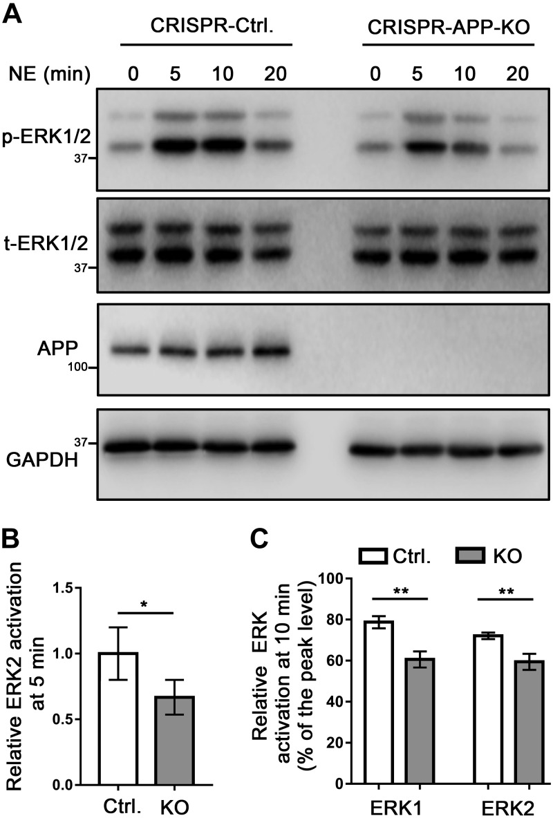
Figure 7.
KO of endogenous APP in N2a cells decreases the strength and duration of α2AAR-mediated ERK signaling. A) Representative Western blots showing p-ERK and t-ERK in control and APP-KO N2a cells. B) Quantification of ERK2 activation with 10 µM NE (plus 1 µM prazosin and 1 µM propranolol) treatment for 5 min. Data (means ± sem) are expressed as fold change vs. that in WT control cells (defined as 1.0). C) Quantification of the relative change in ERK1/2 activation at 10 vs. 5 min of NE treatment. n = 5 in each group. *P < 0.05, **P < 0.01 (Student’s t test).

