Figure 9.
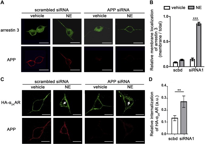
Loss of APP promotes α2AAR-induced arrestin recruitment to the membrane and α2AAR internalization in primary SCG neurons. Primary SCG neurons derived from HA-α2AAR-knock-in mice were transfected with APP siRNA or scrambled siRNA and treated with 10 µM NE (plus 1 µM prazosin and 1 µM propranolol) for 10 min. A) Representative images showing endogenous arrestin 3 and APP. Scale bars, 25 μm. B) Quantification of arrestin membrane recruitment. Data are means ± sem. n = 12 cell per condition. C) Representative images showing internalization of cell surface HA-α2AAR after 10 µM NE (plus 1 µM prazosin and 1 µM propranolol) treatment for 10 min. Arrows: perinuclear punctate staining, which is indicative of endocytosed receptors. Scale bars, 15 μm. D) Quantitation of α2AAR endocytosis. Data are means ± sem (n = 18 cells per condition). **P < 0.01, ***P < 0.001 (Student’s t test).

