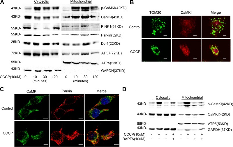
Figure 1.
Mitochondrial depolarization induces the translocation of active pCaMKI, PINK1, and Parkin to the mitochondrion. A) RAW 264.7 cells were exposed to CCCP (10 μM) for indicated times. Total cell and mitochondria were isolated and lysed, and total cytosolic or mitochondrial protein were analyzed by immunoblot (n = 2 independent experiments). B) RAW 264.7 cells were exposed to CCCP (10 μM) for 1 h and analyzed by immunofluorescence (×60 with a 3-fold zoom) for CaMKI and TOM20 (mitochondrial marker; n = 2 independent experiments). C) RAW 264.7 cells were exposed to CCCP (10 μM) for 1 h and analyzed by immunofluorescence (×60 with a 2-fold zoom) for CaMKI and Parkin (n = 4 independent experiments). D) RAW 264.7 cells were exposed to CCCP (10 μM) for 1 h in either the presence or absence of BAPTA [1,2-bis(O-aminophenoxy)ethane-N,N,N′,N′-tetraacetic acid; 10 μM]. Total cell and mitochondria were isolated and lysed, and total cytosolic or mitochondrial protein were analyzed by immunoblot (n = 2 independent experiments). GAPDH, glyceraldehyde 3-phosphate dehydrogenase. Scale bars, 10 μm.

