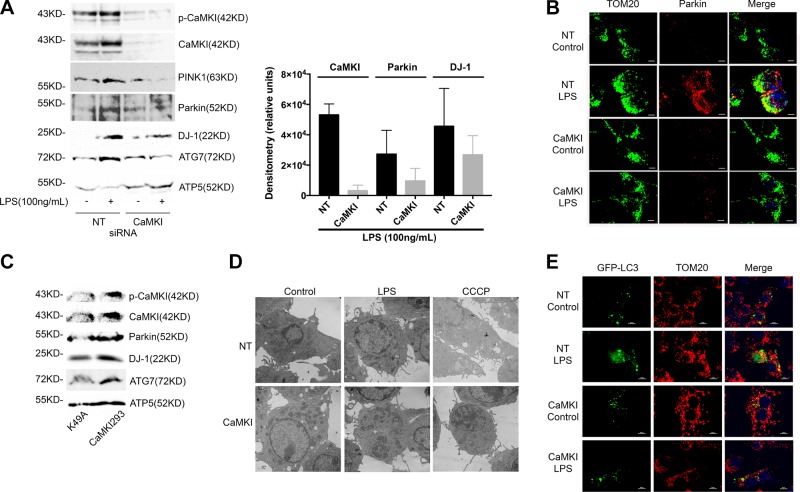
Figure 3.
CaMKI regulates LPS-induced PINK1 and Parkin to the mitochondrion. A) RAW 264.7 cells were transfected with either NT or CaMKI siRNA, and then exposed to LPS (100 ng/ml) for 2 h. Mitochondria were isolated and lysed, and mitochondrial protein was analyzed by immunoblot. Densitometry was performed on CaMKI, Parkin, and DJ-1 using ImageJ (n = 3 independent experiments). B) RAW 264.7 cells were plated on glass coverslips and transfected with either NT or CaMKI siRNA. Cells were exposed to LPS (100 ng/ml) for 4 h, then fixed, permeabilized, and stained with anti-TOM20 or anti-Parkin Ab and analyzed by immunofluorescence (×60 with a 3-fold zoom; n = 2 independent experiments). C) RAW 264.7 cells were transfected with a constitutively active CaMKI (CaMKI293) or a kinase-deficient mutant (K49A) for 8 h. Mitochondria were isolated and lysed, and mitochondrial protein was analyzed by immunoblot (n = 3 independent experiments). D) RAW 264.7 cells were plated on glass coverslips and transfected with either NT or CaMKI siRNA. Cells were then exposed to either LPS (100 ng/ml) for 2 h or CCCP (10 μM) for 1 h. Cells were fixed and analyzed by electron microscopy (n = 4 independent experiments). E) RAW 264.7 cells were plated on glass coverslips and transfected with either NT or CaMKI siRNA. Cells were transfected with GFP-LC3, then exposed to LPS (100 ng/ml) for 2 h. Cells were then fixed, permeabilized, and stained with anti-TOM20 Ab and analyzed by immunofluorescence (×60 with a 3-fold zoom; n = 2 independent experiments). Scale bars, 10 μm.

