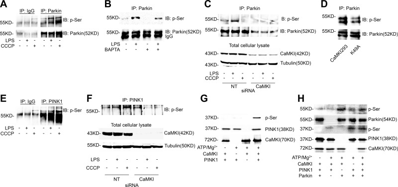
Figure 4.
CaMKI is a PINK1 kinase and regulates Parkin activation. A) RAW 264.7 cells were exposed to either LPS (100 ng/ml) for 2 h or CCCP (10 μM) for 1 h. Total cell lysate was isolated, immunoprecipitated (IP) with IgG control or anti-Parkin Ab, and analyzed by immunoblot (IB) for phosphoserine (p-Ser) or Parkin (n = 2 independent experiments). B) RAW 264.7 cells were exposed to LPS (100 ng/ml) for 2 h in either the presence or absence of BAPTA [1,2-bis(O-aminophenoxy)ethane-N,N,N′,N′-tetraacetic acid; 10 μM]. Total cell lysate was isolated, IP with anti-Parkin Ab, and analyzed by IB for p-Ser or Parkin (n = 2 independent experiments). C) RAW 264.7 cells were subjected to RNAi by using either NT or CaMKI siRNA, then exposed to either LPS (100 ng/ml) for 2 h or CCCP (10 μM) for 1 h. Total cell lysate was isolated, IP for Parkin, and analyzed by IB (n = 2 independent experiments). D) RAW 264.7 cells were transfected with a constitutively active CaMKI (CaMKI293) or a kinase-deficient mutant (K49A) for 8 h. Total cellular lysate was isolated, IP for Parkin, and analyzed by IB (n = 2 independent experiments). E) RAW 264.7 cells were exposed to either LPS (100 ng/ml) for 2 h or CCCP (10 μM) for 1 h. Total cell lysate was isolated, IP with IgG control or anti-PINK1 Ab, and analyzed by IB for p-Ser (n = 2 independent experiments). F) RAW 264.7 cells were subjected to RNAi by using either NT or CaMKI siRNA, then exposed to either LPS (100 ng/ml) for 2 h or CCCP (10 μM) for 1 h. Total cell lysate was isolated, IP for PINK1, and analyzed by IB (n = 2 independent experiments). G, H) One microgram PINK1 (G; MW of recombinant protein, 37.9 kDa) or Parkin (H; MW of recombinant protein, 53.8 kDa) was incubated in the presence or absence of CaMKI (25 ng, MW of recombinant protein, 70 kDa) for 10 min at 30°C with the following additions: 10 mM MgCl2, 0.2 mM ATP, 1 mM CaCl2, and 1 μM CaM. Reactions were terminated by boiling in SDS–2-ME dissociation solution and analyzed by immunoblot (n = 4 independent experiments).

