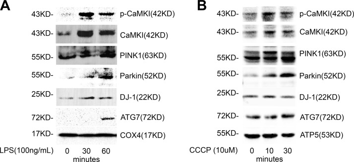
Figure 6.
CaMKI localizes with PINK1/Parkin at the mitochondrion in hepatocytes. A) HuH7 cells were exposed to LPS (1 μg/ml) for indicated times. Mitochondria were isolated and lysed, and mitochondrial protein was analyzed by immunoblot (n = 2 independent experiments). B) Liver harvested from C57BL/6J mice underwent mitochondrial isolation, and isolated mitochondria were exposed to CCCP (10 μM) for the time points shown. Mitochondrial lysate was isolated and analyzed by immunoblot (n = 2 independent experiments).

