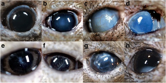
Fig. 1.

Normal and pathologic findings for the anterior segment of the Okarito brown kiwi. Complete ophthalmic examinations consisted of slit lamp biomicroscopy, direct ophthalmoscopy, and streak retinoscopy. Lack of vision was interpreted by no response to light or motion, combined with the severity of ocular lesions (e.g. inability to visualize intraocular structures beyond the abnormal ocular tissue, such as marked corneal or lens opacification). a Normal anterior segment. Note the small palpebral aperture (mean diameter 8.53 ± 0.50 mm SD, n = 9 birds). b Nuclear sclerosis: a normal aging change in the lens associated with changes in lens protein composition. Nuclear sclerosis generally has minimal visual consequences in animals. c Buphthalmia with marked corneal edema. This animal was blind bilaterally but was in good physical condition. d Phthisis bulbi (a globe shrunken with fibrosis). Potential causes include any chronic inflammatory or glaucomatous process or severe trauma. e, f Resorbing, end-stage cataracts. g Anteriorly luxated cataract. h Inferiorly luxated cataract
