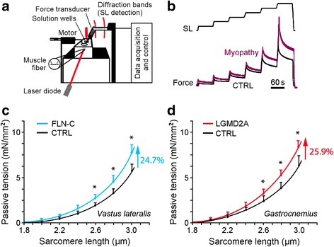
Fig. 1.

Passive sarcomere length-tension relationships of isolated normal and myopathic skinned myofibers. (a) Schematic of experimental setup. (b) Mechanical measurement protocol. Sarcomere length (SL) was increased stepwise from 1.8 to 3.0 μm. (c) Mean SL-dependent passive tension (PT) of normal (CTRL; N = 20 fibers) and MFM-filaminopathy (‘FLN-C’; N = 15 fibers) vastus lateralis muscle fibers, from two different individuals/patients per group. (d) Mean PT of normal (CTRL; N = 14 fibers) and LGMD2A (N = 11 fibers) gastrocnemius muscle fibers, from two different individuals/patients per group. Mean data points were fit with simple exponential functions. Symbols and error bars are means ± SEM; *p < 0.05 in Student’s t-test
