Fig. 3.

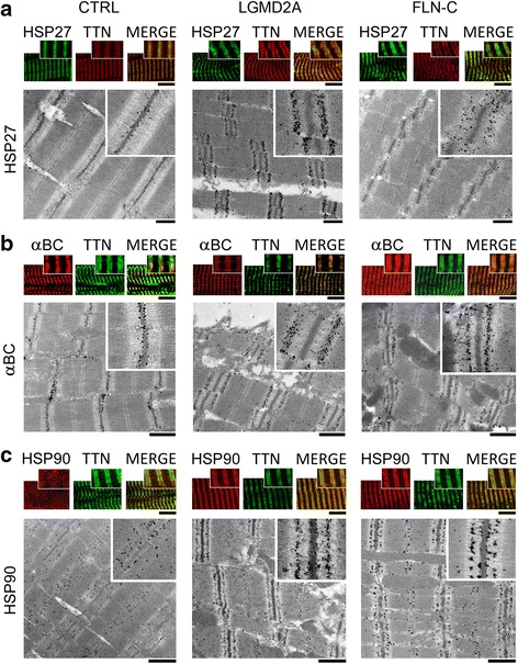
Correlative immunofluorescence and immunoelectron microscopy to localize HSP27, αB-crystallin and HSP90 in normal and diseased myofibers. (a) HSP27 localization on myofibrils. Top panels: representative immunofluorescence images of CTRL, LGMD2A and MFM-filaminopathy (‘FLN-C’) muscle cells labeled with antibodies to HSP27 (secondary antibody: FITC-conjugated IgG). Samples were counterstained against the PEVK titin (TTN) epitope (secondary antibody: Cy3-conjugated IgG); the merged image is on the right in each group. Bottom panels show nanogold-labeled immunoelectron micrographs. Insets, higher-power images of sarcomeric regions immunostained with anti-HSP27/anti-PEVK. (b) Localization of αB-crystallin (αBC). Top panels: immunofluorescence images of myofibers labeled with anti-αB-crystallin (secondary antibody: Cy3-conjugated IgG) and counterstained for PEVK titin (secondary antibody: FITC-conjugated IgG). Bottom panels show immunoelectron micrographs, insets magnifications. (c) Localization of HSP90. Top panels: immunofluorescence images of myofibers labeled with anti-HSP90 (secondary antibody: Cy3-conjugated IgG) and counterstaining for PEVK titin (secondary antibody: FITC-conjugated IgG). Bottom panels show immunoelectron micrographs, insets magnifications. Bars, 5 μm (confocal images) and 1 μm (EM). For a quantitation of nanogold particle distribution on these and similar immunoelectron micrographs, see Additional file 1: Figure S3
