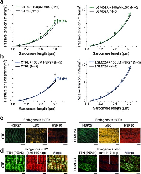
Fig. 6.

Passive tension of skinned normal and myopathy myofibers in the presence of recombinant sHSPs. (a) and (b) Passive sarcomere length-tension relationships of Vastus lateralis muscle fibers from CTRL (left panels) and LGMD2A patients (right panels), before and during incubation with (a) αB-crystallin (αBC) or (b) HSP27 recombinant protein (100 μM). Data points are means ± SEM. The number of fibers measured for each condition (N) is indicated; fibers were obtained from 2 subjects/group. Curves are polynomial fits to the means. *p < 0.05 in Student’s t-test. (c) Localization of endogenous HSP27, αB-crystallin and HSP90 in skinned myofibers after force measurements, monitored by indirect immunofluorescence microscopy. Left panels, CTRL myofibers; right panels, LGMD2A myofibers. (d) Localization of exogenous (6xHIS-tagged) recombinant αB-crystallin, in relation to the PEVK titin epitope (TTN), measured using anti-6xHIS-tag Cy3-conjugated antibodies. Left panels, CTRL myofibers; right panels, LGMD2A myofibers. Insets: Higher-power images of regions-of-interest. Muscle samples were fixed in the stretched state after mechanical measurements and incubated with the respective antibodies. All bars, 5 μm
