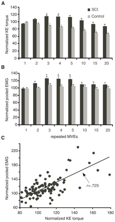
Figure 2. KE Torques and Quadriceps EMG Activity.
Note: Averaged, normalized peak knee extensors (KE) torques (A) and pooled quadriceps electromyographic (EMG) activity (B) during the 1st through 5th, 10th, 15th, and 20th repeated maximal volitional efforts (MVEs) in spinal cord injury (SCI; black) and control (gray) subjects. Asterisks (*) indicate significant increases in torque or EMG during repeated versus baseline MVEs in SCI, or decreases in torque in control subjects. (C) The significant correlation between peak KE torques and pooled EMG for the first 5 repeated MVEs in SCI subjects is also shown (P < .001).

