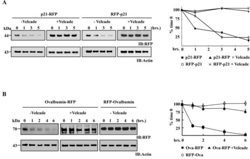
Figure 3. Directionality of proteasomal degradation in living cells.
EL4 cells expressing p21 or ovalbumin N or C-terminally fused to RFP were pre-incubated at 37°C in the presence and absence of 2µM bortezomib (Velcade). After 1 hour cycloheximide was added (50 µg/ml) to the medium for the indicated times. Thereafter, equal numbers of cells were lysed, and the supernatants were subjected to Western blot analyses by anti-RFP and anti-actin antibodies. (A) Cycloheximide chase of EL4 cells expressing p21 fused to RFP either through its C or N-terminus. (B) Cycloheximide chase of EL4 cells expressing ovalbumin fused to RFP either through its C or N-terminus. Quantification of the residual amount of protein detected at each time point during the chycloheximide chase are presented (SD were calculated from three repetitions under similar conditions).

