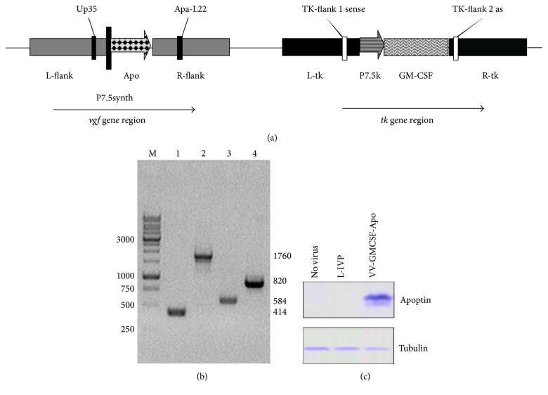
Figure 1.
Verification of recombinant VV-GMCSF-Apo structure and apoptin expression. (a) Schematic view of VV-GMCSF-Apo genome with primer positions indicated. (b) PCR identification of VV DNA and transgene sequences. Electrophoresis of PCR products with primers TK-flank 1 sense and TK-flank 2 as (Lines 1-2) and with primers Up35 and Apa-L22 (Lines 3-4). Lines 1 and 3: wild-type VV (L-IVP); 2 and 4: VV-GMCSF-Apo. M is DNA molecular weight marker. (c) The expression of apoptin was detected by western blot with monoclonal ANTI-FLAG BioM2 antibody. CV-1 cells were infected with VV-GMCSF-Apo or L-IVP for 24 h.

