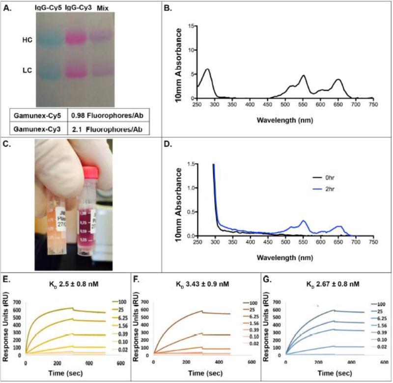
Figure 1. Generation of Cy5- and Cy3-labeled Gamunex-C.
Gamunex-C was labeled with Cy5 and Cy3 and run on SDS PAGE (A). The absorption spectrum for the pre-infusion inoculate containing both Cy5 and Cy3 labeled Gamunex-C was measured with a Nanodrop (B). JM01 Plasma samples at 0hrs and 2hrs post-infusion was assessed both by eye (left, 0hr; right, 2hr) (C) and their spectrum by nanodrop (D). Surface plasmon resonance binding to a CM5 protein A chip for unlabeled Gamunex-C (E), Cy5 labeled Gamunex-C (F), and Cy3 labeled Gamunex-C (G). Antibodies were added in a titration series starting at 100ug/ml and going down to 20ng/ml.

