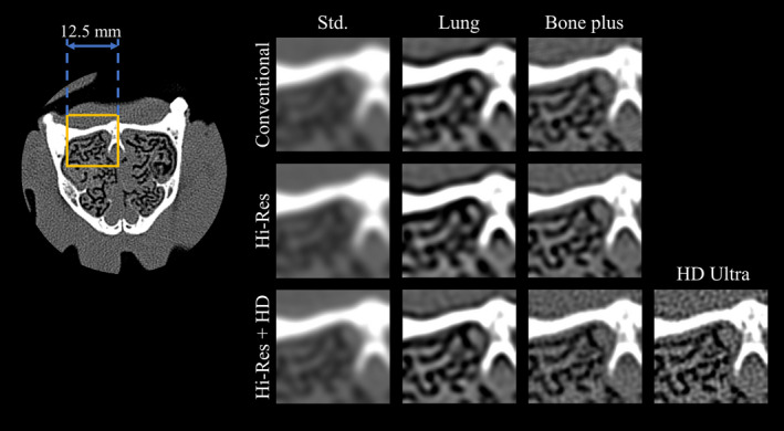
Figure 9.

Axial images of the nasal cavity of the canine subject. ROI images are displayed for both conventional and Hi‐Res modes combined with different kernels. The display window and level are 2000 and 300 HU, respectively. [Color figure can be viewed at wileyonlinelibrary.com]
