
Fig. 5.
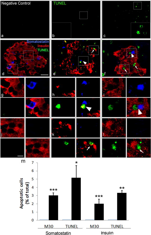
Triple immunofluorescence staining showing apoptotic beta and delta cells in baboons with type 2 diabetes. To reveal the presence of apoptotic cells, a TUNEL assay was performed in pancreas sections from controls (a, b, d, g, j) and two different G4 baboons with type 2 diabetes (c, e, f, h, i, k, l). (a) Negative control (secondary antibodies only); (b, c) TUNEL assay; (d–l) triple immunofluorescence staining with TUNEL (green), insulin (red) and somatostatin (blue). Representative TUNEL-positive somatostatin-labelled cells are shown at high magnification (×2.5) in (g–i). Representative TUNEL-positive insulin-labelled cells are shown at high magnification (×2.5) in (j–l). TUNEL-positive nuclei were detected in a fraction of somatostatin-positive cells (blue, arrowheads) and in insulin-positive cells (red, arrows), only in the pancreas of baboons with type 2 diabetes (a–f; scale bar, 20μm and g–l; scale bar, 10 μm). Quantification of apoptotic delta and beta cells detected using both TUNEL and M30 assay in normal and diabetic baboons is shown in (m). The number of M30-positive and TUNEL-positive delta or beta cells was counted in a total of 100 somatostatin- or insulin-immunoreactive cells in three different baboons for each group. Data are expressed as a means ± SD. *p<0.05; **p<0.01 ; ***p<0.001 type 2 diabetes vs relative control. CTR, control; T2D, type 2 diabetes