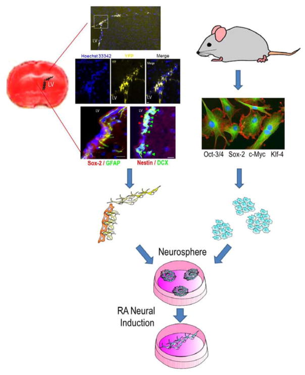
Figure 1. Neural stem cells from the SVZ and induced pluripotent stem cells from somatic cells.
The brain coronal section with TTC staining (red) shows the SVZ region. Immunohistochemical images show multiple markers for neural stem cells in the lateral ventricle SVZ 14 days after an ischemic insult. Using Nestin-Cre-ERT2/Rosa-YFP transgenic mice, the YFP+ cells indicate neural stem cells from the SVZ. Neural progenitors were DCX+ and Nestin+ while neural stem cells were GFAP+ and SOX-2+. Hoechst staining (blue) shows cell nuclei in the lateral ventricle SVZ. These neural stem/progenitors cells can be isolated, cultured and differentiated into neurons. Induced pluripotent stem (iPS) cells can be induced from rodents or humans using a set of reprogramming factors such as Oct-3/4, Sox2, cMyc, and Klf4. iPS cells can also be expanded and differentiated into different cell types including neurons.

