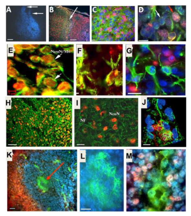
Figure 3. ES cell survival and differentiation after transplantation into the ischemic brain.
Immunohistochemical staining 7 days after intracranial transplantation (14 days post-ischemia). A. Distribution of transplanted ES cells in the post-ischemic basal ganglia revealed by pre-labeled Hoechst (blue) staining. B. Distribution of transplanted ES cells in the post-ischemic cortex. Hoechst-positive ES cells (blue) were present next to undamaged tissue (arrow and the area to the left). NeuN (red) staining shows endogenous neurons in non-ischemic cortical region (left of the arrow) and differentiated neuron-like cells in the ischemic region (right of the arrow). The NeuN staining on the right appears pink in color due to overlap with the blue Hoechst 33342 staining. C. Mouse cell-specific antibodies M2 and M6 (both green) and Hoechst 33342 staining (blue) verified the transplant origin of cells surviving 7 days in the rat host ischemic cortex. NeuN staining (pink) identified differentiated neuronal cells. D. Higher magnification of a confocal image shows the double labeling of Hoechst (blue) and mouse antibody M2/M6 (green) that confirms murine origin of these cells (white arrow). The triple-labeled cells with the additional staining of NeuN (pink) identify ES cell-derived neurons (red arrow). E. Confocal images of NeuN staining (red), mouse antibody M6 staining (green), and overlapped double staining (yellow) show neuronal cells originated from transplanted ES cells in a transplantation area. Scale bar equals 800, 200, 20, 10 and 5 μm, in A, B, C, D and E, respectively. F. and G. Differentiations of transplanted ES cells in the host ischemic cortex and striatum. ES cells were labeled with BrdU or Hoechst 33342 prior to transplantation. The double labeling of BrdU (red) and GFAP (green) identified differentiated astrocytes of ES cell origin (F). An image taken from (G) an 8-μm section shows Hoechst-prelabeled ES cells double stained either with NeuN (red) or GFAP (green), consistent with ES cell differentiation into neurons and astrocytes. Scale bar = 20 μm. H–J. Dendrite growth after ES cell transplantation in the ischemic cortex. NeuN-positive cells and apical dendrite distribution in the ipsilateral cortex. Seven weeks after ES cell transplantation, NeuN and NF double immunostaining in the ischemic core region of the ipsilateral cortex under an inverted fluorescence microscope. Many NeuN-positive cells (yellow or orange) were surrounded by NF labeled processes (green). A confocal image of NeuN/NF-positive ES cell in the 8-Am-thick slice of ischemic cortex. The cell body was positive to NeuN staining (red), cell processes were positive to NF staining (green). The Hoechst labeling (blue) showed several transplanted ES cells. Scale bar = 60 μm in H, 20 μm in I, 10 μm in J. K–M. ES cell-derived Glut-1-positive cells in the ischemic region. Seven days after transplantation, the ischemic core was filled with ES cells (Hoechst-positive, blue) and contained Glut-1-positive vascular-like structures of endothelial cells (green; arrow). Scale bar = 50 μm. Enlarged view of a vascular structure in K (L), positively stained with Glut-1 (green). Scale bar = 10 Am. Glut-1-positive endothelial cells (green) were also co-labeled with Hoechst (blue), verifying their origination from transplanted ES cells (M). The pink color is from neighboring NeuN-positive cells. Images were taken using an inverted fluorescence microscope. Adopted from Figures in Wei et al., 2005 in compliance with the journal’s copyright policy.

