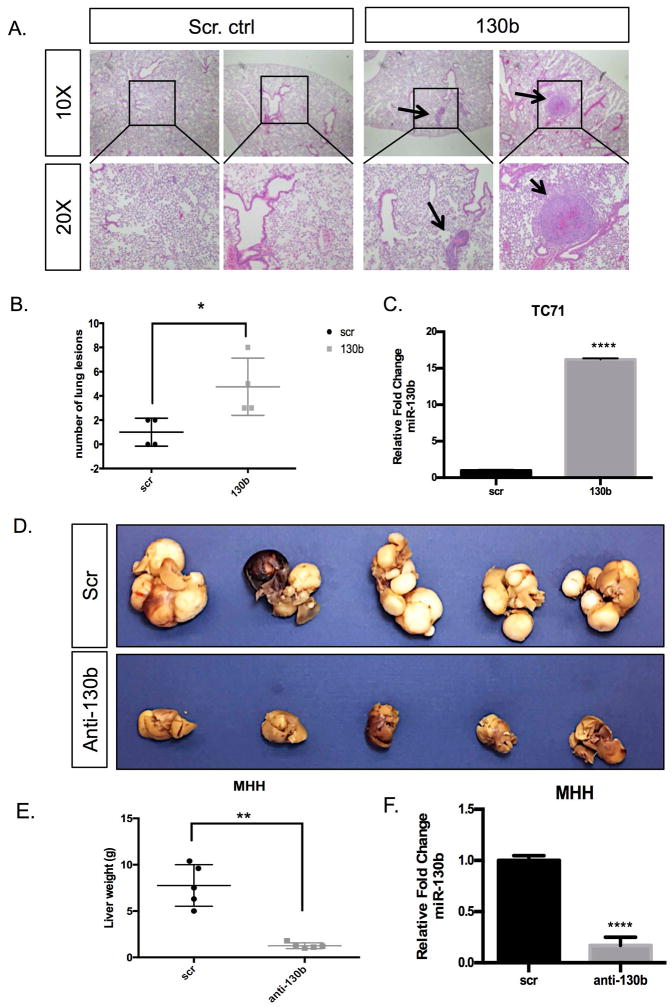
Figure 2. miR-130b alters metastatic potential in vivo.
(A) H&E-stained lung sections from the control and miR-130b expressing groups following tail vein injections of 1 × 10⁶ TC71 cells with and without stable overexpression of miR-130b. Black arrows highlight evident microscopic lesions. (B) Quantification of pulmonary nodules in each group. Error bars were generated from control (n=5) and miR-130b (n=4) in each group. *P<0.05 (C) qPCR expression of miR-130b in TC71/scr and TC71/130b stable cell lines. (D) Images of metastatic livers of scrambled control and anti-miR-130b stable expressing groups following tail vein injections of 1 × 10⁶ MHH cells with and without knockdown of miR-130b. (E) Quantification of miR-130b expression in MHH/scr and MHH/anti-130b stable cell lines. Error bars were generated from control (n=5) and anti-miR-130b (n=5) in each group. **P<0.05. Black boxes indicate areas magnified and arrows represent metastatic lesion observed.

