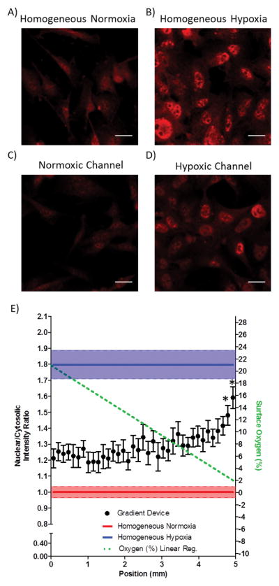
Figure 4.

HIF-1α activation in endothelial cells occurs at low oxygen levels within a linear oxygen gradient. (A–B) Panels of HIF-1α immunofluorescent staining for normoxic and hypoxic homogeneous control devices. (C–D) HIF-1α immunofluorescent staining in the oxygen gradient device of cells directly above the normoxic gas supply microchannel and hypoxic gas supply microchannel, respectively. Scale bar: 20 μm. (E) Quantification of the nuclear to cytosolic ratio as a function of the position in the oxygen gradient. * p < 0.05 as compared to homogeneous normoxia.
