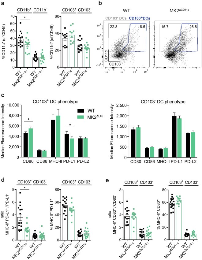
Figure 3.
MK2ΔCD11c mice accumulate stimulatory PD-L1− CD103-expressing DCs. (a) CD11b and CD103-expressing DCs within tumour-infiltrating CD45+ leukocytes as measured by flow cytometry. (b) Representative plots showing gating strategy of CD103+ and CD103− DCs. Cells were pre-gated for live, single, CD45+ CD11c+ cells. Frequency within parental population is indicated. (c) Surface receptor expression on intratumoural CD103+ and CD103− DCs. (d) Ratios of PD-L1+/PD-L1− cells within MHC-II+ DCs and frequency of MHC-II+ PD-L1+ cells within the indicated parental DC population. (e) Ratios of CD80+/CD80− cells within MHC-II+ DCs and frequency of MHC-II+ CD80+ cells within the indicated parental DC population. Each symbol represents one individual animal. Data are presented as mean ± SEM and pooled from two to three independent experiments (n = 5–8 mice per group each). *P < 0.05. P-values were determined using Student’s t-test.

