Fig. 3.
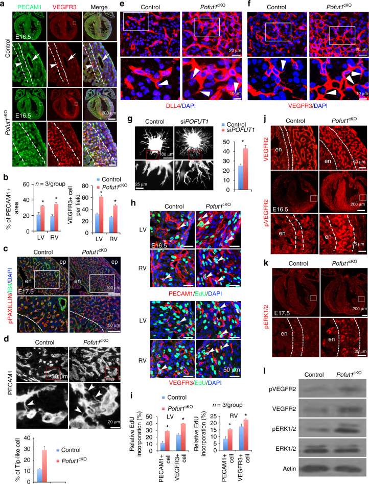
POFUT1 regulates angiogenic functions and proliferation of coronary angiogenic cells. a, b PECAM1/VEGFR3 co-staining of E16.5 heart sections shows VEGFR3high coronary angiogenic cells (arrowhead) in the inner myocardium (between dotted lines) and VEGFR3low coronary arteries (arrow). b Quantification of PECAM1+ areas and the number of VEGFR3high cells. *p < 0.01. c IB4 and pPAXILLIN co-staining of E17.5 heart sections. en/ep, endocardium/epicardium. d PECAM1 immunostaining showing increased tip-like cells (arrowhead) in myocardium of E16.5 Pofut1 cKO embryos. *p < 0.01. e, f Immunostaining of DLL4 and VEGFR3. g Spheroid sprouting assay with phalloidin staining showing increased sprouts by inhibition of POFUT1 using shRNA. Eight spheroids per group, n = 3, *p < 0.001. h, i EdU labeling for proliferative PECAM1+ or VEGFR3+ cells (arrowheads). *p < 0.05. All bar charts represent mean ± SD. j immunostaining of VEGFR2 and pVEGFR2. k Immunostaining of pERK1/2. l Western blot analysis of pVEGFR, VEGFR2, pERK1/2, and ERK1/2 expression in E16.5 control and Pofut1 cKO hearts

