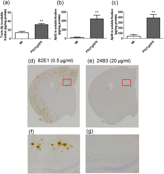
Figure 2.
24B3 detects the toxic conformer of Aβ42 in the soluble fraction from the brain of AD model mice. (a–c) The levels of the toxic conformer of Aβ42 (a), total Aβ42 (b), and total Aβ40 (c) in the soluble fraction of the brain of PS2Tg2576 at 6 months of age. (d–g) Immunohistochemical staining of brain sections from PS2Tg2576 was performed with 82E1 (0.5 µg/ml) (d and f), and 24B3 (20 µg/ml) (e and g). The slices were exposed to formic acid for antigen activation. High magnification images of the area inside the red rectangles (d and e) are shown in f and g. **p < 0.01.

