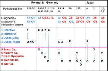
Table 3.
Esophagus Case 1: distribution and combination patterns of diagnoses: biopsy (B1: X )–ESD2 (O), Poland: 1–20, Germany: G1–3, Japan: J1–6
This table shows distribution and combination “patterns” of diagnoses
Reactive/Regenerative reactive lesion or regenerative lesion, Carcinoma (SM~) carcinoma with invasion to submucosal or deeper layer, Dyspl. (Low) low grade dysplasia, Dyspl. (High) high grade dysplasia, Susp. Ca suspicious of carcinoma, Noninv. Ca noninvasive carcinoma, X-O represents just “combinationpattern” of diagnoses between biopsy (X) and corresponding ESD (O) specimens, but not the “exact number” of the combination itself

