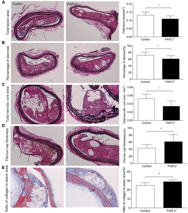
FIGURE 1.

Morphometric analysis data. Representative images of EVG-stained brachiocephalic arteries exhibited significantly reduced total lesion area (A), reduced percentage of stenosis (B), reduced total necrotic core area (C) and increased fibrous cap thickness (D) in PAR-2-/- mice. Representative images of Masson staining within brachiocephalic arteries showed significantly increased plaque collagen content (E) in PAR-2-/- mice, as compared to the controls. Data represent mean ± SD. ∗P < 0.05.
